
- at 4 to 6 gestational weeks, the hip joint develops from the cartilaginous anlage;
- by 7 weeks a cleft develops between precartilagenous cells which are programmed to form the femoral head and acetabulum;
- by 11 weeks, the hip joint formation is largely complete;
- femoral head is completely encircled by the acetabular cartilage;
- at late gestation, femoral head grows more rapidly than acetabular cartilage, so that at birth femoral head is less than 50% covered;
- at birth, acetabulum is at its most shallow and most lax inorder to maximize hip ROM which facilitates the delivery process;
- hip is uncontained in extension and adduction reflecting hip shallowness;
- after several weeks, acetabular cartilage develops faster than the femoral head, which allows progressively more coverage;
- normal occurance of hip shallowness and capsular laxity in the neonatal period are inital factors factors involved in DDH;
- references:
- Growth and development of the acetabulum in the normal child. Anatomical, histological, and roentgenographic studies.
- The fetal acetabulum. A histomorphometric study of acetabular anteversion and femoral head coverage.
- Morphometric study of the fetal development of the human hip joint: significance for congenital hip disease.
- Histological study of the fetal development of the human acetabulum and labrum: significance in congenital hip disease.
- Growth characteristics of the fetal ligament of the head of femur: significance in congenital hip disease.
- Acetabular Anteversion with CT in Supine, Simulated Standing, and Sitting Positions in a THA Patient Population
- Femoral Anteversion: 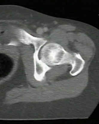
- femoral version is defined as the angular difference between axis of femoral neck and transcondylar axis of the knee;
- normal values:
- on average, femoral anteversion ranges from 30-40 deg at birth and decreases progressively throughout growth
to about 15 deg at skeletal maturation;
- in most adults, anteversion averages between 8 and 14 deg, w/ an average of 8 degrees in men and
14 degrees in women;
- note however, wide variation of femoral anteversion seen in multiple studies from 10-15 deg
retroversion to 30 deg anteversion;
- exam:
- medial rotation of thigh in extension exceeding 70 deg is abnormal;
- Reider Test:
- prominence of the greater trochanter indicates axis of the femoral neck;
- noting the position of the patella, an accurate determination of the femoral anteversion can be determined;
- references:
- Femoral anteversion. A necessary angle or an evolutionary vestige?
-
Femoral anteversion in the hip: comparison of measurement by computed tomography, magnetic resonance imaging, and physical examination.
- Femoral anteversion
- Relationship between femoral anteversion and findings in hips with femoroacetabular impingement
- Femoral version of the general population: does "normal" vary by gender or ethnicity?
- Increased Anteversion of Press-fit Femoral Stems Compared With Anatomic Femur
- Femoral Version Abnormalities Significantly Outweigh Effect of Cam Impingement on Hip Internal Rotation
- Acetabular Anteversion: (see optimal acetabular anteversion for THR)
- references:
- Acetabular inclination and anteversion in infants using 3D MR imaging.
Femoral Version Abnormalities Significantly Outweigh Effect of Cam Impingement on Hip Internal Rotation
Clinical determination of femoral anteversion. A comparison with established techniques.
Intertrochanteric versus supracondylar osteotomy for severe femoral anteversion.
Medial femoral torsion and osteoarthritis.
Lower-extremity rotational problems in children. Normal values to guide management.
The anatomy and functional axes of the femur.
Femoral anteversion and physical performance in adolescent and adult life.
The Prevalence of Acetabular Retroversion Among Various Disorders of the Hip.
Ischial Spine Projection into the Pelvis. A New Sign for Acetabular Retroversion