
Discussion
- indicated for symptomatic soft tissue impingement, synovitis:
- specific lesions amenable to arthroscopic debridement include:
- osteochondral lesions;
- meniscoid lesion in anterolateral gutter;
- mass of fibrocartilagenous tissue arising from the tibio-fibular joint will protrude into the joint;
- patients will note anterolateral ankle pain, popping, and giving way;
- anterior impingement snydrome of the ankle
- thickening of antero-inferior tibiofibular ligament;
- arthrofibrosis following ankle fracture;
Technical Considerations
- use 30 deg wide angle - 2.7 mm arthroscope (if not available, then use 4 mm scope);
- consider use of a pump set for a pressure of 50 mm;
- use 3.5 mm shaver;
Modern ankle arthroscopy was popularised by amongst others James Guhl in the United States in the eighties and is now a common place intervention in most Foot and Ankle surgeons repertoires.
Many of the pathologies which cause symptoms in the ankle are soft tissue in origin and poorly imaged by MRI. A normal scan should definitely not be a reason not to intervene in a joint that has failed to recover and which has well localised and congruent symptoms. Ankle arthroscopy has a significant role in the treatment of the post traumatic joint (after fracture or severe sprain) as well as early ankle arthritis from any cause.
There are a variety of portals described for ankle arthroscopy but the vast majority of pathology in almost every intra-articular location in my experience can be dealt with through the basic antero-lateral and antero-medial portals described in this technique. The keys are having appropriate portal placement and a wide enough range of appropriately sized (and angled) instruments. Posterior ankle arthroscopy is a different beast for different indications and demonstrated by Nick Cullen from Stanmore elsewhere on OrthOracle at Posterior Ankle decompression-Arthroscopic technique . A comprehensive review of the range of other portals is to be found in the paper by Ferkel & Scranton (Current concepts review:Arthroscopy of the Foot & Ankle. J Bone Joint Surg.1993. 75-A:1233-1242. R.D.Ferkel , P.E.Scranton) or any of James Guhls instructional works.
Instantaneous recovery never occurs after an ankle arthroscopy despite instantaneous removal of the causative pathologies. The joint linings need to heal just as any operated soft tissues and this may take 6-12 weeks, condition dependent. Full recovery after microfracture of osteochondral defects is longer again in most cases by another couple of months.
The set up and detailed technique described here is basically the one taught to me by Paul Cooke during my fellowship in Oxford fifteen years ago. My guess is he worked most of the tricks out himself, but some may just originate from elsewhere.
Watch the full instructional video for this technique by subscribing.
This overview is brought to you by Orthoracle - the online e-learning Orthopeadic Surgery Atlas
View this procedure on OrthOracle.com | OrthOracle is a commercial partner of Data trace LLC
mechanical distraction
- some sort of mechanical distraction device is useful;
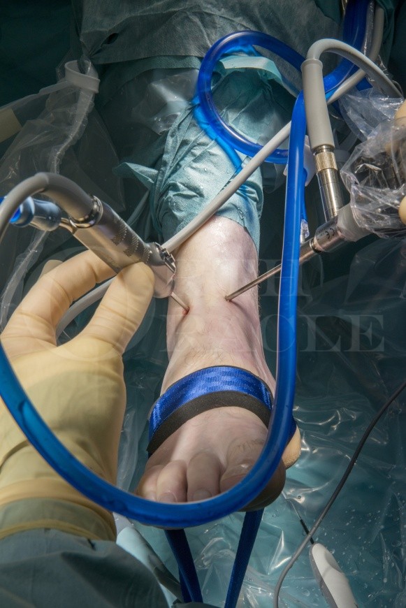
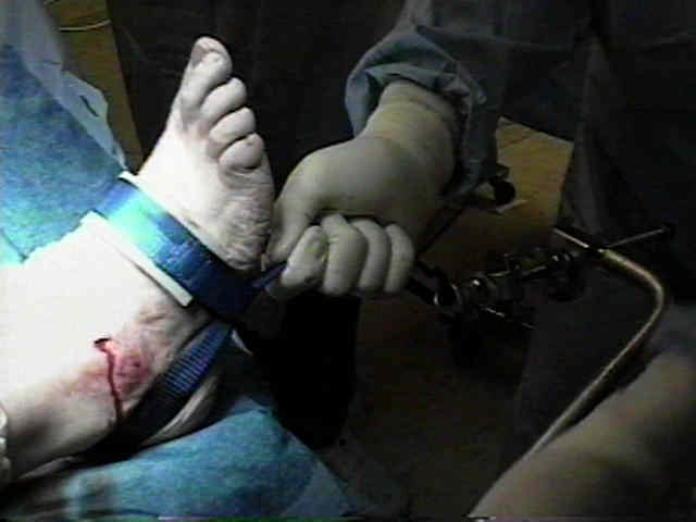
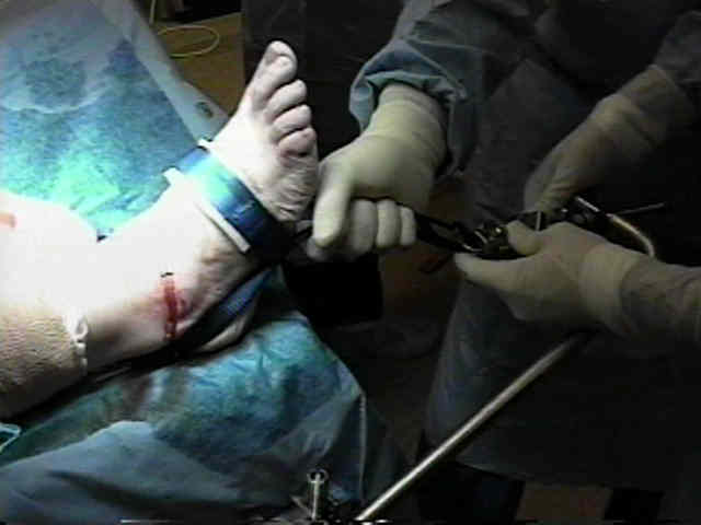
- typically the kit will contain sterile straps which are applied to a sterile metal bar after the leg is prepped;
- usually a sterile strap is wrapped over the dorsum of the foot and heel;
- usually about 25 lbs of distraction force is required, which gives between 1 to 1.5 mm of distraction;
cautions
- distraction of more than 30 lbs for more than 1 hour is associated with reversible nerve conduction changes;
- excssive use of force for prolonged periods of time may cause bothersome paresthesias in the superficial peroneal nerve;
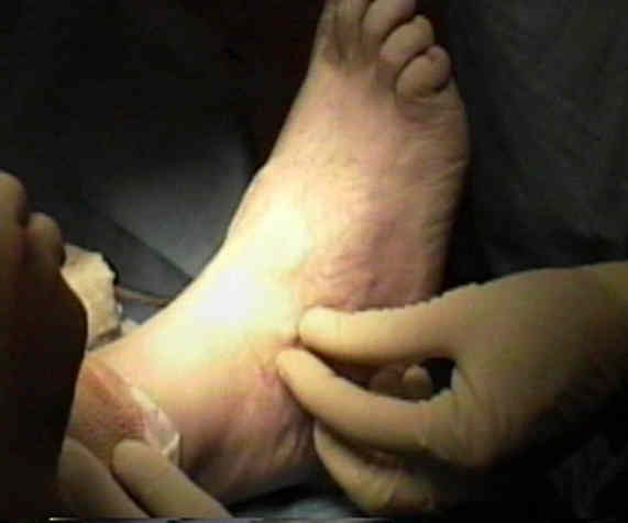
Position
- patient is supine with leg placed in a arthroscopic leg holder (as for knee scopes);
- leg hangs free so that knee is flexed to about 90 deg;
- a sterile kerlex cloth band is wraped in figure of 8 fashio around the foot and ankle, with the free end tied into a loop which is then positioned just above the floor;
- surgeon's foot is placed in the kerlex loop and is used to distract the ankle joint;
- reference: A simple distraction technique technique for ankle arthroscopy.
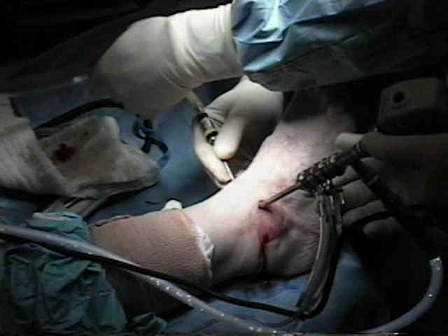
Portals
- use a 15 blade to carefully incise thru skin only;
anteromedial
- initial arthroscopy is performed with the scope in the anteromedial portal, but for the majority of case, this portal will be used for instrumentation;
- located at the level of the ankle joint, just medial to the tibialis anterior tendon, and located about 5 mm proximal to the medial malleolus;
- 18 gauge syringe is used to infuse saline into the joint;
- greater saphenous nerve and vein are at risk w/ this portal, lying 7-9 mm medial to the portal;
anterolateral
- once joint is distended w/ saline, use 18 gauge needle to mark location of anterolateral portal which should lie just lateral to peroneus tertius tendon;
- staying lateral to the peroneus tertius, helps avoid injury to the dorsal lateral branch of the peroneal nerve;
- use the scope to transilluminate the anterolateral skin, inorder to look for underlying cutaneous nerves;
- scope can then be driven forward (elevating the synovium and skin) which further assists with placement of this portal;
- make small incision and then spread w/ hemostat;
- be aware that the intermediate branch of the superficial peroneal nerve is about 5-6 mm from this portal;
- reference Anatomic relations between ankle arthroscopic portal sites and the superficial peroneal and saphenous nerves.


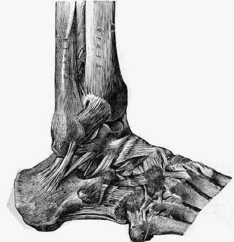
Sequential Examination
- visualization from the anteromedial portal:
- deltoid ligament;
- medial malleolus;
- medial gutter (medial talomalleolar joint);
- talar dome (osteochondral lesions)
- anterior gutter;
- tibiofibular joint:
- synovitis, fibrocartilagenous protrusion;
- posterior tib-fib ligament;
- anterior tib-fib ligament;
- anterior talofibular ligament (arising from the tip of the fibula)
References
- Current Concepts Review. Arthroscopy of the Ankle and Foot.
- Diagnostic and operative arthroscopy of the ankle. An experimental approach.
- Arthroscopic treatment of synovial impingement of the ankle.
- Arthroscopic treatment of anterolateral impingement of the ankle.
- Noninvasive ankle distraction: relationship between force magnitude of distraction, and nerve conduction abnormalities.