
Discussion
- acute arthritis caused by CPPD crystal-induced inflammation;
- almost as common as gout & may perfectly mimic gout during acute flare;
- pseudogout attacks occurring before age 50 are uncommon;
- see: pseudogout occurence after joint replacement
- references:
differential diagnosis
- pseudogout occurence after joint replacement:
- septic arthritis:
- pseudogout may be confused with septic arthritis;
- chondrocalcinosis of the meniscus occurs not only in otherwise healthy individuals in older age groups but also in definite association w/ several distinct metabolic disorders;
- references:
- trauma:
- perhaps the majority of cases of chondrocalcinosis occur from trauma;
- reference: Localized chondrocalcinosis in traumatized joints.
- hemochromatosis;
- hyperparathyroidism (most common);
- up to 30% of hyperparathyroid pts have chondrocalcinosis;
- hypothyroidism;
- gout:
- reference: Gout and coexisting pseudogout in the knee joint.
- hyperparathyroidism
- hypothyroidism
- hemochromatosis
- ochronosis
- ochronosis is a hereditary enzyme deficiency (homogentisic acid oxidase) resulting in deposition of homogentisic acid polymers in articular cartilage
- acromegaly
- Paget's disease;
- hypomagnesemia
Clinical Features
- most often affects the knee and the wrists;
CPPD Crystal Exam
- Crystal Examination of Synovial Fluid:
- Calcium pyrophosphate dihydrate crystals are visualized under compensated polarized light microscopy
- crystals may be more difficult to detect than MSU crystals because of their smaller size, more intraphagolysosomal location, & less brilliant colors;
- in contrast to MSU crystals, CPPD crystals show weak positive birefringency and have squared or rhomboidal shaped ends;
- aggregates do not show birefringence (or are weakly birefringent) under polarized light;
- alizarin red stain, can confirm that these clumps are masses of calcium crystals;
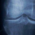
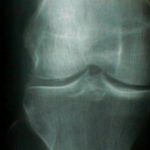
Radiographic Analysis
- punctate and linear densities in hyaline or fibrocartilage, which are found in knee menisci, acetabular labrum, & TFCC;
Therapeutic Principles
- aspiration of joint and steroid injection, once diagnosis of infection has been excluded, will usually control symptoms;
- indomethacin;
- colchicine? may be useful for pseudogout;
- magnesium on an as needed basis
- arthroscopic lavage: